Definition of sleep
Simple behavioral definition: The body loses a reversible behavior of perception and response to the surrounding environment and is a complex physiological and behavioral process;
Divided by status: NREM & REM sleep;
EEG performance of EEG: NREM sleep EEG performance synchronized slow wave, waveform characteristic spindle wave, K-conformity wave and high amplitude slow wave, stage (I, II, III),
REM sleep EEG activity is active, activation, muscle tension is slow, accompanied by rapid movement of the eye;
Central and peripheral nervous system REM phase: pons-knee-occipital POG wave, accompanied by eye movement, distal muscle twitching, middle ear muscle activity; spinal motor neurons are affected
Inhibition, muscle relaxation, fluctuations in cardiopulmonary activity, accompanied by dreams.
Start of sleep
The definition of sleep initiation is identified from the state of physiological indicators
EMG
Electrooculogram
EEG
Behavior accompanying sleep
Simple behavioral tasks (cognition & driving);
Visual response (spark stimulation is suppressed);
Auditory response (expiration or disappearance of stimulation latency);
Olfactory response (latency is extended or disappeared, but special odors can be sniffed during any sleep period);
Response to meaningful stimuli
Sleep muscle myoclonus
Memory before sleep begins
(A meaningful stimulus usually leads to a K-complex or a wake-up, with a relatively low threshold sensitivity)
(The cerebral palsy area is activated when a meaningful sound is stimulated; the bilateral sacral frontal cortex is activated when there is no meaningful stimulation)
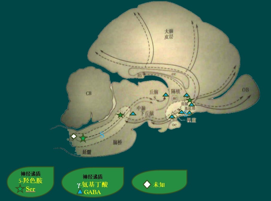
Sleep-wake-related nervous system
Identification of brainstem sleep production system
The dorsal medullary reticular formation and solitary tract nucleus neurons can induce slow wave activity in the cortex under awakening state and produce sleep;
It may be caused by inhibiting the neurons of the upstream reticulating system, and may also directly affect the forebrain system;
The solitary tract nucleus receives the introduction of the glossopharyngeal nerve and the vagus nerve (the 9th and 10th branches of the cranial nerve), and the baroreceptors and chemoreceptors from the visceral organs of the chest and abdomen;
The solitary nucleus and the efferent nerve of the dorsal stenosis with the reticular structure project upward to the pons and the midbrain, many of which terminate in the parabrachial nucleus;
The parabrachial nucleus projects to the thalamus, hypothalamus, preoptic area, amygdala and frontotemporal cortex, which belong to the visceral-marginal forebrain;
The solitary tract nucleus not only inhibits reticular agonism, but also the forebrain marginal system associated with sleep production.
Forebrain sleep production system
The anterior hypothalamic - basal forebrain and preoptic area, and the lower brainstem are important areas for inducing sleep;
The production of cortical spindle waves in the cortex is necessary, but it is not necessary for the generation of cortical slow waves and sleep behavior;
The orbitofrontal cortex and the basal forebrain, preoptic area, and anterior hypothalamic neurons together constitute the sleep induction system of the forebrain.
Single cell recording sleep-activated neurons
Only a few neurons in the brain increase their frequency of distribution during slow wave sleep, and are concentrated in the brainstem;
There are also some neurons in the solitary tract nucleus that are more active than the awake state during slow wave sleep. These sleep-related neurons coexist with high-frequency spontaneous discharge neurons in the region when they wake up;
Relative to the arousal state, the frequency of discharge in the hypothalamic anterior, preoptic and amygdala neurons increased during slow wave sleep;
In slow wave sleep, the discharge activity first appears as the spindle wave of the reticular nucleus of the thalamus, 12-14 Hz, consistent with the thalamus-cortex projection frequency;
The intrinsic properties of thalamic and cortical projection neurons also appear as slow-wave 0.1-4 Hz slow waves at other frequencies. This slow wave activity is also projected from the cortex to the striatum, basal forebrain, brainstem, spinal cord, and thalamus, and is enhanced by the central nervous system to play an important role in slow wave sleep.
Chemical substances (neurotransmitters or neuromodulators) in slow wave sleep
★ 5-hydroxytryptamine
5-HT can attenuate cortical activity and promote slow wave sleep production by acting on different receptors of different cell types;
5-HT inhibits cholinergic neurons in the pons, midbrain and basal forebrain;
5-HT stops discharging during heterophasic sleep, further confirming that it is not a key substance for maintaining sleep; biochemical studies have released 5-HT in REM & NREM sleep
Less than the arousal state, it is proved that 5-HT neurons are not very important in sleep maintenance, nor are they necessary for slow wave sleep.
★Adenosine
Adenosine acts as a neuromodulator that inhibits neuronal firing through a second messenger-coupled post-synaptic receptor, which also acts on other receptors to prevent nerve endings from releasing
Transmitter
In the thalamic cortex, adenosine hyperpolarized projection neurons promote neuronal burst discharges of slow wave activity in slow wave sleep. So adenosine is in the brain like in the periphery
The same function is exerted, and when the cells continue to move, accumulation can prevent the cells from being excessively active and damaged;
Adenosine in the brain is associated with a specific burst discharge of the thalamic-cortical circuit, which forms the basis of slow wave sleep;
Accumulation of adenosine outside the brain (1 molecule of ATP) may be a factor inducing brain fatigue and sleep.
[ATP cannot be stored because ATP must be consumed in a short time after synthesis. Adenosine triphosphate (ATP) is a nucleotide that acts as a "molecular currency" for intracellular energy transfer, storing and transferring chemical energy. ATP also plays an important role in nucleic acid synthesis. Adenosine is an endogenous nucleoside distributed throughout human cells. It can directly enter the myocardium to phosphorylate to form adenosine, participate in myocardial energy metabolism, and also participate in the expansion of coronary vessels and increase blood flow. Adenosine has a physiological effect on the cardiovascular system and many other systems and tissues of the body. Adenosine is an important intermediate for the synthesis of adenosine triphosphate (ATP), adenine, adenylate, and adenosine. 】
★ γ-aminobutyric acid
GABA is the main inhibitory neurotransmitter in the brain and is of great significance in sleep;
In the brainstem network, relatively small GABAergic neurons are mixed with larger amino acid neurons, and glutamatergic neurons of the ascending reticular agonistic system are inhibited by local projection;
Thalamic reticular neurons containing GABA produce thalamic-cortical spindle waves and inhibit the successor neurons of the thalamic cortex through inhibitory postsynaptic potentials. It inhibits the cerebral cortical afferent pathway-thalamic, which is the basis for slow wave sleep and loss of consciousness;
GABA neurons are distributed in the anterior hypothalamic-anterior anterior and basal forebrain and descending to the brain region of the posterior hypothalamus, inhibiting arousal system neurons;
GABAA is directly linked to the chloride channel and acts rapidly; GABAB acts through the second messenger system to regulate different potassium and calcium channels, which are slow and long-lasting;
Both are involved in the super-correlation associated with slow waves and spindle waves. Participated in the basic induction and maintenance of slow wave sleep, first by inhibiting the arousal system, and secondly by inducing, regulating and stabilizing the spindle wave and slow wave activity based on the thalamic-cortical explosive discharge.
★Cerebrospinal fluid factors, peptides and slow wave sleep
Enkephalin, endorphin, dynorphin in endogenous opioid peptides, associated with sensory regulation and analgesia, may play a role in the initiation and maintenance of sleep;
Somatostatin mainly inhibits central nervous cells;
Cortistatin is structurally and physiologically similar to somatostatin;
Growth hormone releasing factor;
Prostate D2 (mainly glial cell synthesis) has a circadian rhythm consistent with the sleep-wake cycle.
★ blood-borne factors and slow wave sleep
5-HT is a platelet produced and present in the blood, promoting sleep production in specific areas of the brain outside the cerebral blood barrier;
The accumulation of insulin during awakening leads to the production of sleep, also to the periventricular organs and nearby nuclei outside the blood-brain barrier, especially the basal inferior brain, the solitary tract nucleus and the vagus nerve region;
Cholecystokinin is an enteric hormone secreted after ingestion and inhibits further food intake. This anorexic hormone also promotes rest and induces slow wave sleep;
The intestinal hormone bombesin is also released after ingestion, forming a feeling of satiety, indirectly through the vagus nerve into the orphan nucleus or directly acting on the periventricular organs outside the virtual brain barrier, inducing rest and sleep after meals;
The muramyl peptide substance is synthesized by intestinal bacteria and has a function of inducing sleep. Like other bacteria and viruses, it promotes the synthesis and release of interleukins and other cytokines and induces slow wave sleep;
When the body is stimulated by the outside world, hormones or peptides released from the intestines or the immune system can cause sleep;
Therefore, like awakening regulation, the influence of blood-derived substances on the central sleep system, partly through the periventricular organs to the brain, these factors may also be certain pituitary hormones, such as growth hormone, prolactin, they are in slow wave sleep Released, so it may also have an effect on sleep.
EEG activity and sensory treatment during sleep
basic concept
★High amplitude and synchronized EEG rhythm-----distinguish between SWS, NREM, REM sleep and awakening
★Awakening and REM sleep period----activation ("de-synchronization")
Means high amplitude and synchronous brain wave interruption, high frequency and low amplitude instead of low frequency oscillation wave
Synchronized oscillation of the thalamic cortex system and the cellular basis of the activation pattern
★ NREM time low frequency oscillation of the thalamic cortex network
Spindle oscillation; δ oscillation (1-4Hz); slow oscillation (‹1Hz);
★ brainstem--thalamic circuit participates in arousal and REM brain electrical activity and fast oscillation (20-50Hz)
Sensory information processing
★ Excitability enhancement
★ Abrasion and brain activation during REM sleep
★ Selective attention: The general arousal effect can be divided into two parts: selective attention and movement start;
P100 may prompt confirmation of the input signal, P300 reflects the non-specific decision and closes;
★ Alert and deep phase event path in REM sleep
There are two types of neural pathways that may be involved in the phase process induced by external sensory stimulation during the arousal phase, and phase events that are stimulated by brain storage information during the REM sleep phase;
(A pathway consists of GABGergic neurons of the substantia nigra SNr (black matter network) and its distribution in the hypothalamus, the upper arm paraventricular area and the thalamic nucleus of the brainstem reticular formation, and finally through the thalamus to the pons The nucleus of the eye; in addition, the cholinergic parabrachial nucleus (or pons nucleus) in the junction of the substantia nigra and the midbrain-ponsal junction is also connected by nerve projection.)
An alert stimulus must be able to use two completely different systems. The first system: the SNr-thalamic-ponsal pathway, which is responsible for the phase shift of gaze involved in localization; the second system consists of the paraventricular stem region and the thalamic projection target of SNr; pons - geniculate-occipital region (ponto – geniculo - occipital PGO ) waves, PGO waves are widely distributed in the corresponding cortical regions outside the thalamic nucleus and visual cortex; hypothalamic and forebrain structures (which are thought to store some emotional information) may generate PGO for driving brainstem neurons It is the most effective.
★ Modification suppression (guarantee the selection of the input signal and the adjustment of the output signal, necessary to maintain the adaptive state. Ach GABA EEG synchronous oscillation activity basis)
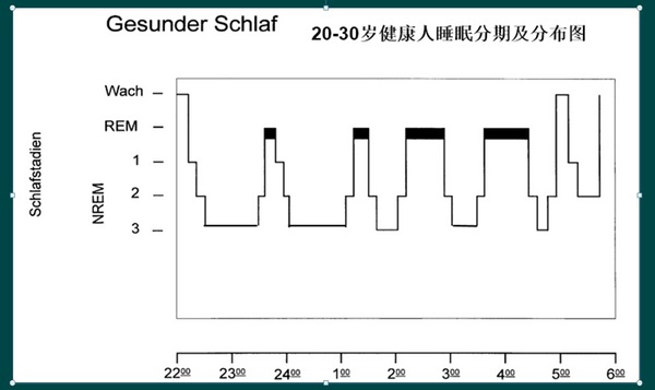
Healthy adult sleep structure overnight sleep process
1. Entering NREM sleep from waking up;
2, NREM-REM sleep cycle duration is generally 90 minutes;
3, one third of the night before sleep is mainly SWS, related to sleep initiation;
4, one third of the night after REM-based, and the body temperature circadian rhythm;
5, NREM sleep usually accounts for 75% -80%;
6, REM sleep accounts for 20%-25% of total sleep, appearing 4-6 times per night;
REM sleep (REM)
Clear and vivid "dream"
The EEG amplitude is reduced similarly to the arousal, the new cortex exhibits a low voltage, and the hippocampus presents a regular high voltage θ wave.
Slow muscle tone;
Genital erection, expansion changes;
The body temperature regulation disappears and the body temperature fluctuates with the ambient temperature;
The lateral geniculate PGO wave maps to the thalamic nucleus and the neocortex (ponto – geniculo - occipital PGO);
Irregular heart rate; (irregular activity is not controlled by a single pacemaker)
Produces peripheral or middle ear muscle twitching signals that may precede or be secondary to PGO and rapid eye movement
REM generation mechanism
★ disconnect research
The pons is a key part of REM sleep and is the result of dynamic interaction between the forebrain and brainstem mechanisms. Deletion of muscle tone requires activation of the motor inhibition system in the spinal cord.
★ Damage research
Deletion of the under-blue nucleus
★Stimulation research
Cholinergic agonists stimulate the strongest form of REM sleep (injected into specific parts of the ventral blue plaque of the pons)
★ neuron activity
REM-distribution cells (REM-priming): the neurotransmitters that release the cell population are gamma-aminobutyric acid, acetylcholine, glutamic acid or glycine;
REM-quiet cells (REM-off): the neurotransmitters that release the cell population are norepinephrine, adrenaline, serotonin and histamine;
Muscle tension control
During REM sleep, inhibition of motor output is accompanied by inhibition of sensory transmission, and both have similar neurological mechanisms.
REM sleep function
Evolutionary explanation: The inactivation state of NREM leads to slower metabolism and lower response. Arousing from REM sleep is more alert than NREM.
REM rebound phenomenon: during REM sleep, histamine, norepinephrine and serotonin neurons are terminated, and this termination does not occur in the awake state, so waking is not a substitute for REM sleep function. Rebound may be the accumulation of demand for monoaminergic cell population passivation. When the release is reduced, the synthesis of monoamines and receptors is accelerated, and agonists are not required, and the receptors of these substances can be desensitized. But this hypothesis is not fully supported.
The Oxford File Holder is designed with digital printing, silk screen printing, UV printing and other printing methods and accessories. It is a combination of practicality and aesthetic culture.
Jilin Y.F. Imp & Exp Co., Ltd is an exporter and manufacturer (Cang nan Y.F. Stationery & Gift Co., Ltd.)in Creative products, such as Backpack ,Shoulder Bag, Pencil Case , Handbag,Multifunctional Bag. Coin Purse .Cosmetic Bag.Storage bags. File holder.Canvas handbag and Notebook etc. which is a professional stationery company setting research and development, producing, sales and trade into one. Our company always takes quality, service, efficiency and innovation as our management philosophy. Since our brand Y.F. has been put on the market, the products sell well throughout the country consistently, and be exported to Europe and America,, and South America countries as well as regions, where the product enjoys great customer loyalty and good population. Choose Y.F. is not to choose a batch of stationery, but to choose a commitment and responsibility, Thanks for your attention, support, trust an cooperation. Wish to establish long-term business relationship with you in the near future.


Oxford File Holder,File Holder Box,File Folder Organizer Rack,Wall File Holder
Jilin Y.F. Import & Export Co.,Ltd , https://www.jlpencilcase.com